The Back Part of the Eye Where Transduction Occurs
It must first pass through other layers that process it so that it can be interpreted by the retina. But light does not impinge on the retina unaltered.
17 5 Vision Concepts Of Biology 1st Canadian Edition
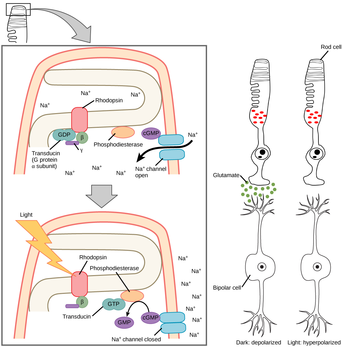
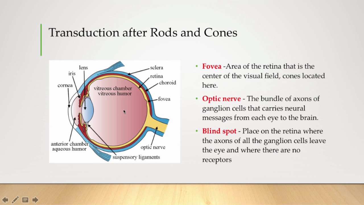
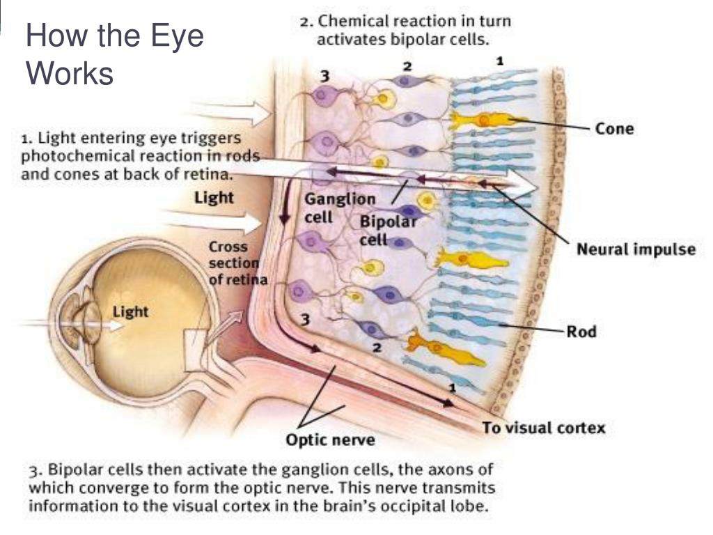
The rods and cones are the site of transduction of light to a neural signal.

. However light does not enter the retina unaltered. Cells of the retina. Circular sheet of cells that line the back of the eyeball and contain a photo pigment known as a chemical that absorbs light.
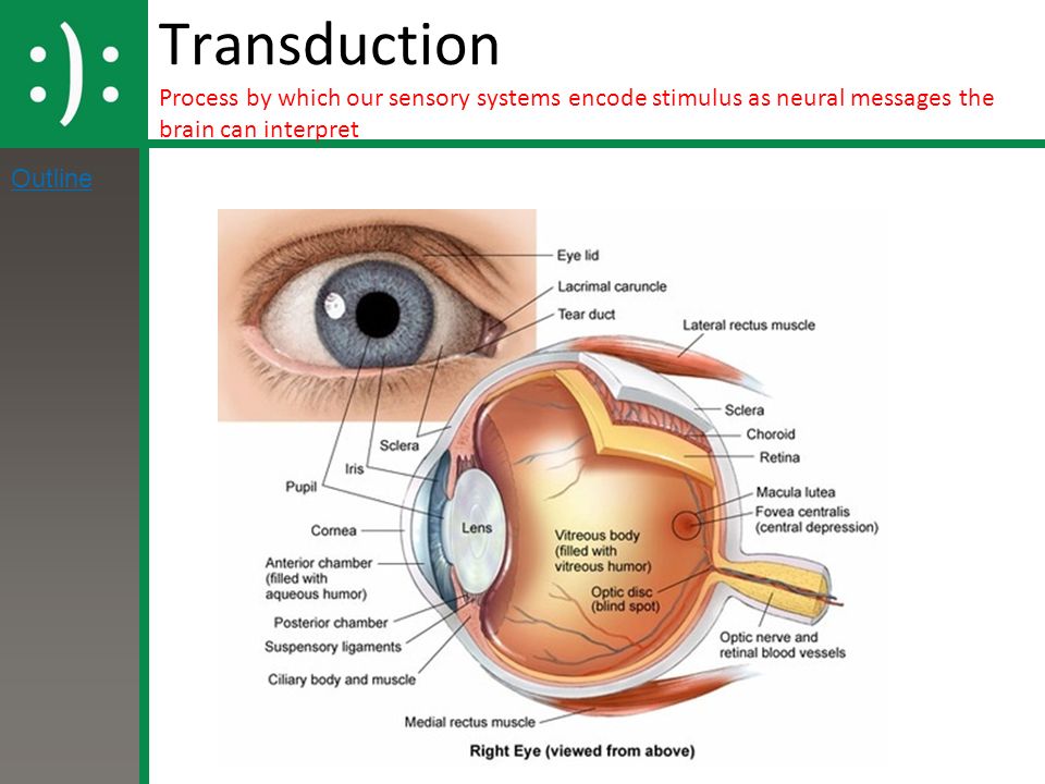
The retina a thin layer of cells located on the inner surface of the back of the eye consists of photoreceptive cells which are responsible for the transduction of light into nervous impulses. The Inner Ear is usually referred to as the Cochlea in dark green in the picture on the next screen. However light does not enter the retina unaltered.
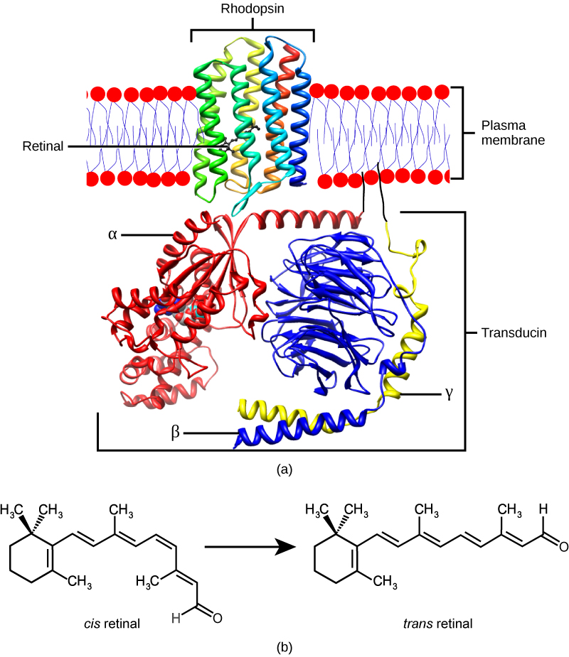
Electromagnetic Spectrum what we can see. In vertebrates the main photopigment rhodopsin has two main parts Figure 1. Eye too short far away objects focus behind retina usually ok near objects cannot compensate with focusing power.
In what part of the eye does transduction occur. It passes through other layers that process it so that it can be interpreted by the retina see figure b below. Most of the process occurs on the back of the eye called the retina.
Asked Sep 25 2015 in Anatomy Physiology by Naynadine. The Inner Ear Cochlea is where transduction takes place. Both rods and cones contain photopigments.
An opsin which is a membrane protein in the form of a cluster of α-helices that span the membrane and retinala molecule that absorbs light. It is basically a tube filled with fluid. It must first pass through other layers that process it so that it can be interpreted by the retina.
The retina is the most important part of our eye it is often referred to as the brain of the eye. Uneven curvature of eye prevents proper focusing of light. GTPase activity is involved in the regulation of signal transduction because it.
The function of the Outer and Middle ear was to conduct sound energy to the Inner Ear where the actual transduction takes place. It is made up of several layers of cells and the light must pass through all of them to experience transduction. Transduction begins in the in the backmost layer of the retina made up of cells called photoreceptors setting off a chain reaction in response to light.
The retina a thin layer of cells located on the inner surface of the back of the eye consists of photoreceptive cells which are responsible for the transduction of light into nervous impulses. The first step in the visual transduction process that occurs in the retina is. There are two structures of the eye which help to focus light on the retina.
Asked May 6 2021 in Biology Microbiology by MadDecent. However light does not enter the retina unaltered. What ear structure vibrates back and forth when sound waves strike.
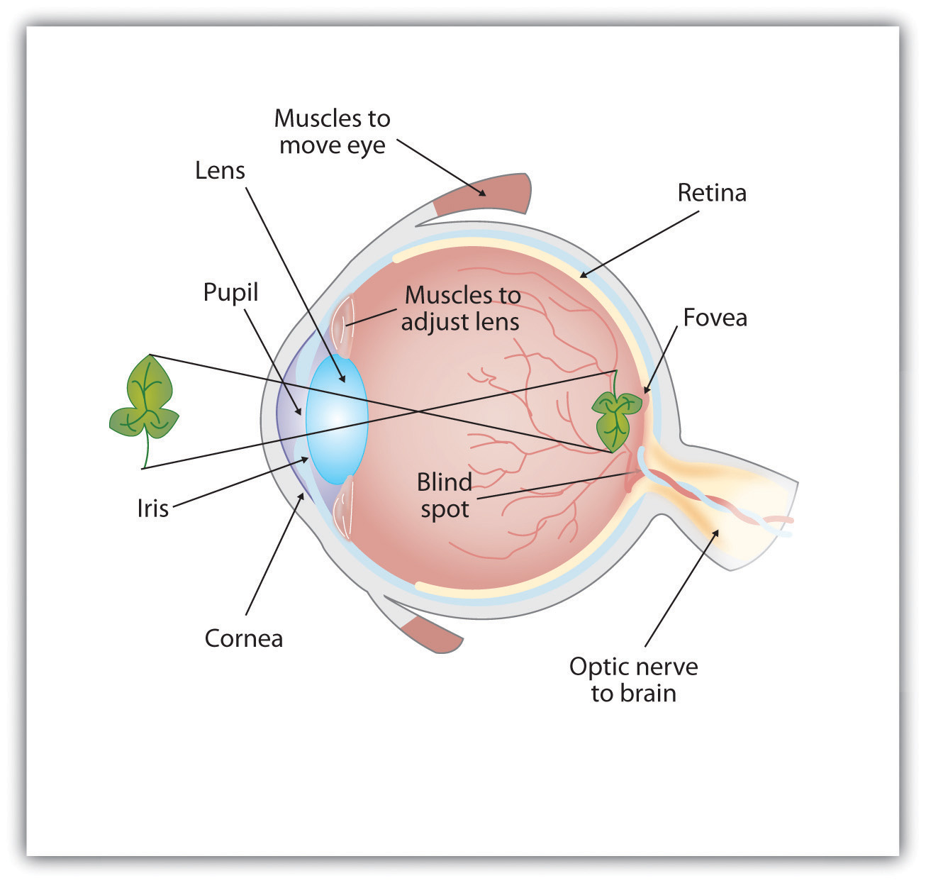
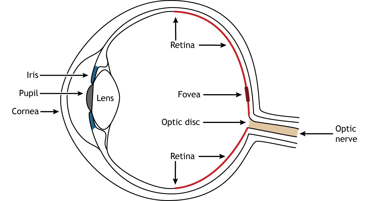
The photoreceptive cells of the eye where transduction of light to nervous impulses occurs are located in the retina shown in the figure below on the inner surface of the back of the eye. It must first pass through other layers that process it so that it can be interpreted by the retina. The cochlea is to the EAR as the RETINA is.
What part of the eye does transduction occur. - Visible spectrum for humans ranges from 400 to 700 nanometers. The first step in the visual transduction process that occurs in the retina is.
The cochlea is to the as the is to the eye for transduction. First it is important to know that the retina is made up of several layers of cells and the light must pass through all of them to experience transduction kind of like a water filtration process. First neural component of visual transduction located at the back of the eye adjacent to the optic nerve.
The retina a thin layer of cells located on the inner surface of the back of the eye consists of photoreceptive cells which are responsible for the transduction of light into nervous impulses. The retina is the most important part of our eye. Dynamic equilibrium is maintained by the.
Anatomy of the Eye. The retina a thin layer of cells located on the inner surface of the back of the eye consists of photoreceptive cells which are responsible for the transduction of light into nervous impulses. The lens reflects the light upside down and inverted toward the back of the eye which is retina.
Occurs in the fundus back layer of the retina.

Visual Transduction Sensory Transduction The Nervous System Medical Physiology 3rd Edition
![]()
The Signal Transduction Cascade In Rods And Rod Bipolar Cells The Download Scientific Diagram

Vision Biology Ii
5 2 Seeing Introduction To Psychology 1st Canadian Edition

5 2 Seeing Introduction To Psychology 1st Canadian Edition
![]()
Unit 4 Sensation Perception And States Of Consciousness Ppt Video Online Download

Anatomy Of The Eye Biology For Majors Ii
17 5 Vision Concepts Of Biology 1st Canadian Edition
Vision The Retina Foundations Of Neuroscience
![]()
5 Vision Cognitive Neuroscience David Eagleman Jonathan Downar Ppt Download

Structure And Function Of The Eye Vision Mcat Content

Sight Sensation And Perception Katelyn Rodgers Ap Psychology Magazine
![]()
Kristen Erin Jyra Michelle Andrew And Kayla Ppt Video Online Download
Visual Processing Part 2 Transduction Youtube
Jhs Ap Psychology Unit 4 Sensation Perception Essential Task 4 2 Describe The Sensory Process Of Vision Including The Specific Nature Of Energy Transduction Ppt Download

Anatomy Of The Human Eye And The Retina With Its Specialized Cells 1 Download Scientific Diagram
![]()
Light Reception By Eye And Signal Transduction To Scn Rods And Cones Download Scientific Diagram

Sensory Processes Boundless Psychology
Ppt Sensation And Perception Chapter 4 Powerpoint Presentation Free Download Id 1837106